What is ultrasound anyway?
Ultrasound refers to the use of ultra-high frequency (> 20 kHz) sound waves to create images based on the echoes that are received after reflecting off body structures.
Transducers
The most common types of ultrasound transducers or probes are the curvilinear (left), phased array (middle), and linear probe (right).
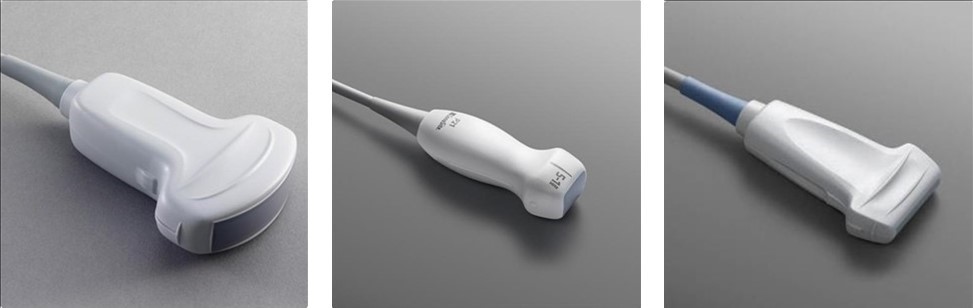
The curvilinear probe is a low frequency probe (2-5 MHz) which is generally used for visualizing deep structures such as abdominal organs. The phased array (2-7.5 MHz) is optimal for visualizing moving objects and therefore is the go-to for cardiac imaging. The linear probe is a high a frequency probe (7.5-11 MHz) meaning it is ideal for visualizing superficial structures such as skin, soft tissue, nerves, and blood vessels.
Describing an Image
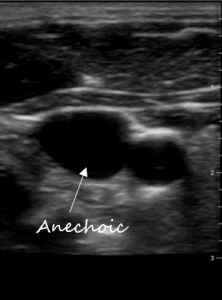
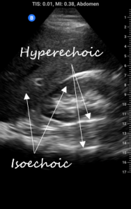
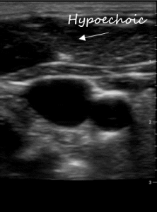
Communication works best when were are all using the same language. Describing ultrasound images is no different. All ultrasound terms are based on describing how sound is reflected. Below are terms used to describe the appearance of body structures on ultrasound:
Basic knobology
For purposes of basic POCUS we will consider just 2 functions that are consistent across all ultrasound devices: depth and gain. We will also review 3 modes: brightfield (B-mode), motion-mode (M-mode), and colour doppler.
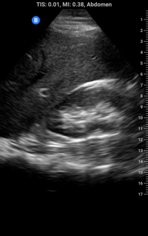
Depth: increasing depth brings far away structures onto the screen and simultaneously makes everything appear smaller; think of it like zooming out on digital map, you will seem more but with less detail. Decreasing depth does the opposite, we don’t seem as far down but there is greater detail. Optimal depth is achieved when all structures of interest are in the image field but as large as possible. Watch what happens as we increase depth in the images below (note increasing depth markers on the right of each image):

Gain: this can be thought of as the ‘brightness’. The higher the gain the whiter things get; the lower the gain the darker things get. Gain is a processing function and doesn’t change any of the characteristics of the ultrasound beam itself. Optimal gain is achieved when anechoic structures like fluid or blood are black and hypoechoic tissue like subcutaneous tissue or organs are grey and hyperechoic structures like arterial walls, pleura, and bone are white.

B-mode: Brightness mode, or B-mode, is the standard mode for ultrasound images and shows a 2D greyscale image.
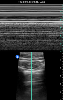
M-mode: Motion-mode or M-mode is another common image mode. M-mode creates an image by plotting the signal from one single ultrasound beam over time. It is used commonly in cardiac ultrasound for showing the movement of heart valves or visualizing lung sliding.
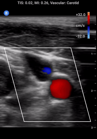
Colour doppler: in this mode a small window appears on the screen that detects the velocity and direction of flow which is represented using colour. At the side of the screen a colour scale will appear; the colour on top represents flow towards the probe and colour on the bottom away from the probe.
Scanning Orientation
Even though POCUS is primarily used to make decisions at the bedside it is important to make sure that images can be interpreted afterwards by anyone. It is therefore critical that we all scan the same way.
As a rule we always position the machine and perform scans on the patient’s right, just as we would a physical exam.
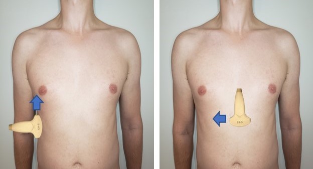
There are also 2 standard orientations to all scans: longitudinal with the probe marker towards the patient’s head parallel with the body from head to toe (shown below on the right), and transverse with probe marker to the patient’s right making an axial cut (left).
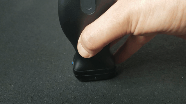
The 5 Key Movements
The language of probe movements similar to orientation and image description uses a standardized language. The 5 cardinal movements of ultrasound are demonstrated in the clips below.

Sliding: Moving the transducer without altering the longitudinal or transverse orientation.

Tilting: Angling the beam along a fixed longitudinal plane.

Rotating: Keeping the vertical axis fixed and twisting the probe clockwise or counterclockwise.
Rocking: Or heel toeing, angling the beam along a fixed transverse plane.

Compressing: Applying firm downward pressure.
